Fig. 4.

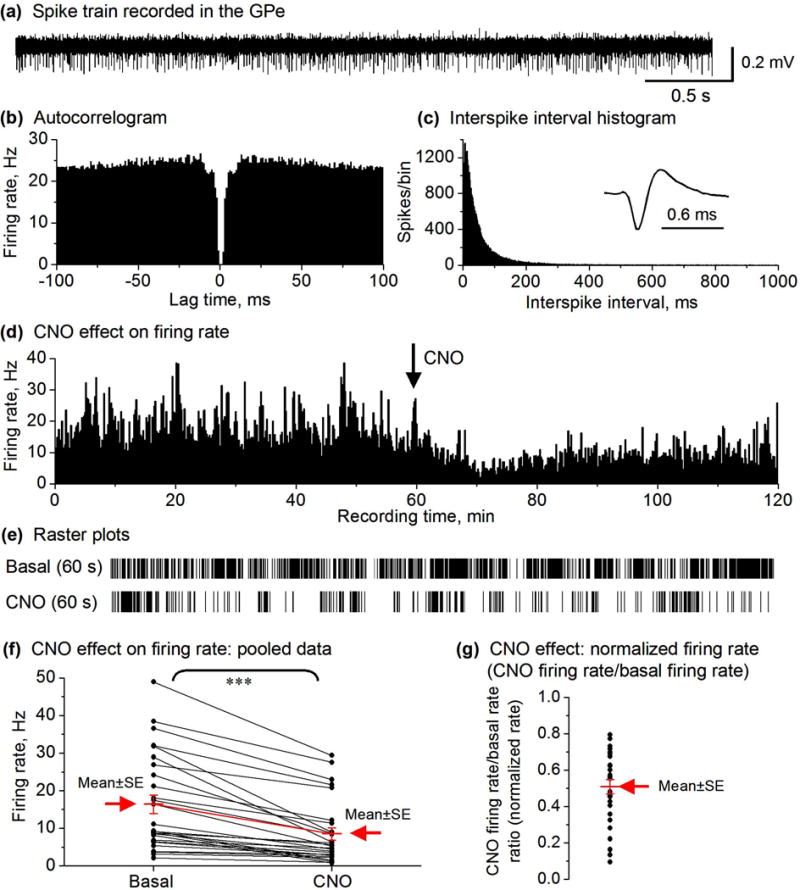
CNO IP injection decreased the spike firing in the majority of GPe neurons (27 out of total 33) in D2-MSN Gs-DREADD mice. (a) Representative example of spike train recorded in the GPe in a DREADD positive mouse. (b,c) autocorrelogram and ISI histogram (1 ms bin width) characterizing a GPe neuron recorded in DREADD positive mouse. (d) Spike firing rate histogram of a GPe neuron recorded in DREADD positive mouse showing a decrease in the firing rate after 2.5 mg/kg CNO injection. Firing rates were calculated and plotted as the average frequency of discharge in 10-s bins. (e) Raster plots showing the spikes in a representative 60 s before and after CNO injection of an isolated GPe neuron. (f) Paired before-after scatter plot showing that CNO decreased the firing rate of the 27 GPe neurons in D2 Gs-DREADD positive mice (n=3), *** p<0.001, paired t-test. (g) CNO reduced the normalized firing rate in GPe neurons. Each black dot represents the normalized firing rate of a single GPe neuron. The mean ± SE are shown in red and indicated by the arrow.
