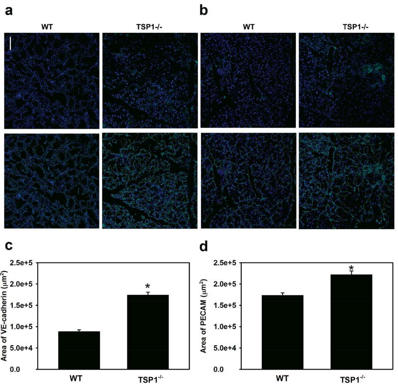
Fig. 3. Area of VE-Cadherin and PECAM Is Decreased in Lacrimal Glands in TSP-1−/− Compared to WT Mice.
Female WT and TSP-1−/− mouse lacrimal glands were removed at 12 weeks of age, fixed, and stained with VE-cadherin (a) and PECAM (b) antibodies to study blood vessels. Representative micrographs are shown in a and b. Quantification of positive staining was performed and the total area was calculated. Data are mean ± SEM from four independent animals and shown in c and d. * indicates a significant difference from WT..

