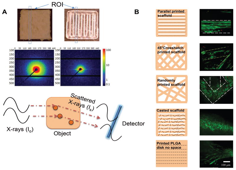
Figure 1. 3D printing induced polymer molecule alignment and resulted cell alignment.
A. SAXS set up and results comparing casted and printed PLGA scaffolds. Scattered X-ray waves arrive at the detector at different time to form signals as a result of material intrinsic structure. X-ray scattering shows uniform scattering in all directions of the casted scaffold, which indicates random polymer molecule organization. However, scattering shows higher intensity on the horizontal direction of the printed scaffold, which indicates vertically oriented polymer molecule existing after printing. Blue boxes indicate regions of interest (ROI) upon incoming x-ray. B. Cell alignment on scaffolds printed with different patterns. Confocal microscope image showed hMSCs aligned differently on printed patterned scaffolds. Live cells were shown in green and dead cells were shown in red. Short black lines indicates the polymer molecule alignment.

