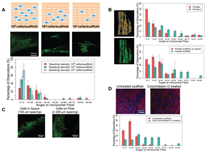
Figure 2. Cell alignment quantification and the impact of different factors on cell attachment.
A. Cell alignment and quantification for different seeding densities. Cells seeded at various concentration all showed aligned pattern, with more than 80% of the total population had an angle to the fiber less than 30 degree. B. Cell alignment quantification for scaffolds with different patterned and fabrication methods from Figure 1. Individual cells were selected manually in ImageJ, then the angles were automatically calculated by the built in Plugin OrientationJ. Cells on scaffolds with parallel pattern mostly aligned along the fibers, by displaying an alignment angle to the printed fiber of less than 30°. As a comparison, cells on random printed fibers aligned with less order, displaying more evenly distributed angles. As a control, cells on casted scaffold showed evenly distribution of attachment angles. In a nother control using printed solid disk without any patterns, cells showed directional alignment along the printed fibers. All cells from the captured images were included for analysis. C. Preferred adhesion with different scaffold spacing. With a 200 μm printed fiber, when the width of the spacing is smaller than the fiber, most cells appeared in the gap spacing rather than attached on the surface of the scaffold. However, when the spacing was larger than the diameter of the fiber, cells tended to attach on the scaffold surface instead. D. Phalloidin staining of actin fibers and comparison of cell alignment before and after cytochalasin D treatment. After cytochalasin D treatment, the attached cells completely lost their organized direction due to loss of focal adhesion.

