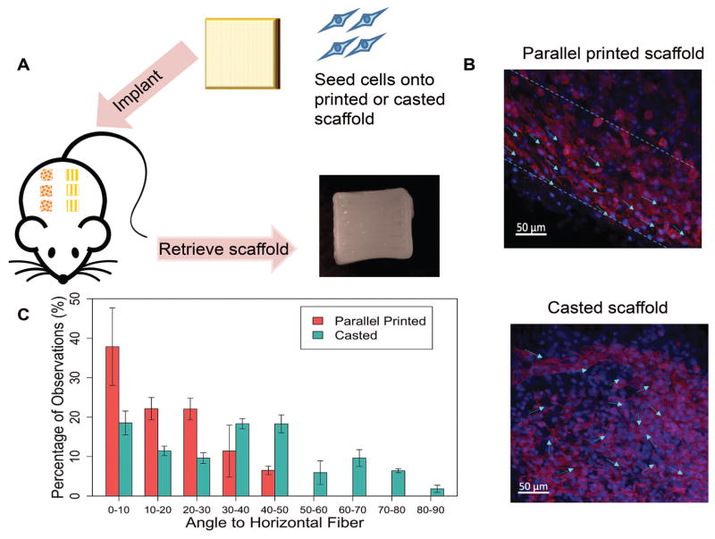
Figure 3. In vivo cell alignment evaluation and quantification.
A. Experimental flow of the animal study. Rat MSCs were seeded on parallel printed or casted scaffolds, then scaffolds were implanted subcutaneously in rats. After 7 days, retrieved scaffolds showed intact structure and printed patterns. B. Cell alignment was maintained after in vivo implantation. Cells appeared mostly aligned on the parallel patterned scaffold, while cells were more randomly distributed on the casted scaffolds. C. Quantification of cell alignment in vivo. The average number was calculated from three different images.

