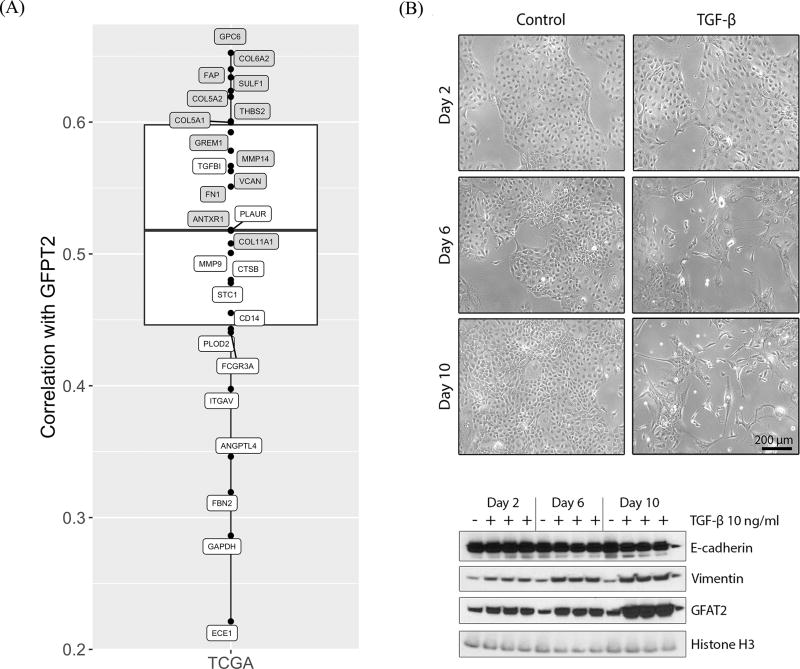
Figure 4.
Validation of association of GFPT2 and SUVmax-associated secreted glycoproteins and EMT. (A) Correlation between GFPT2 and glycoprotein-coding genes correlated with glucose uptake in TCGA AD. Genes highlighted in gray were more expressed in CAFs compared to other cells in our TME cohort; these genes are among highest correlated. (B) Morphological and protein expression changes in HCC827 cells after EMT induction with TGF-β treatment. (Top) Phase-contrast microscopy showing HCC827 cells after treatment with, or without (control), TGF-β (10 ng/ml) up to 10 days. All images were obtained at a magnification of 100×. Scale bar represents 200 μm. (Bottom) Following TGF-β treatment on HCC827, protein lysates were harvested at the indicated time points and E-cadherin, Vimentin and Glutamine fructose-6-phosphate amidotransferase 2 (GFAT2: protein coded by GFPT2 gene) were analyzed by Western blot. Histone H3 was used an internal loading control. During the EMT time course, Vimentin increased, E-cadherin decreased and GFAT2 increased.

