
Fig. 2.
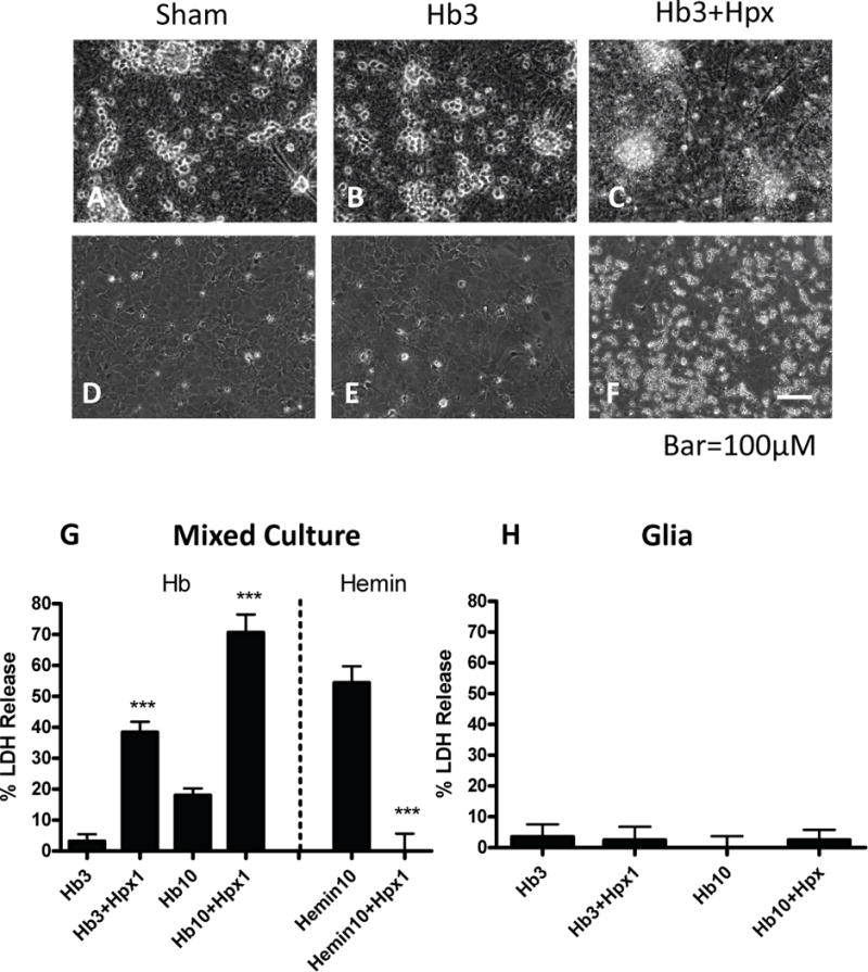
Effects of hemopexin on hemoglobin and hemin toxicity. (A-C) Phase contrast images of mixed neuron-glia cultures subjected to: (A) sham medium exchange only; phase bright neuron cell bodies cluster in groups and overlie a confluent glial monolayer; B) hemoglobin (Hb) 3 μM for 24 h; (C) Hb 3 μM plus hemopexin (Hpx) 1 mg/ml; neuronal degeneration and a fine precipitate are apparent, while the glial monolayer remains intact. (D-F) Glial-only cultures treated as in A-C; glial monolayer remains intact; precipitate is apparent in Hb+Hpx condition. (G) Bars represent mean neuronal loss (± S.E.M., n = 7 cultures for Hb 3 μM condition, 15 cultures/condition for Hb 10 μM, and 8 cultures/condition for hemin experiments) in mixed neuron-glia cultures as measured by LDH assay. (H) Percentage glial cell loss (± S.E.M., n = 12 cultures/condition) in glia-only cultures treated with Hb alone or with 1 mg/ml Hpx as in G. The low LDH values in sham cultures were subtracted from each mean value to yield the signal associated with neurotoxicity. ***p < 0.001 versus value in Hb or hemin only groups.