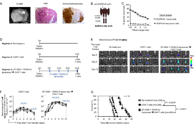
Fig. 7. Nanoparticle preconditioning improves CAR-T cell therapy of glioblastoma.

(A) T2 MRI scan, H&E and immunohistochemical analysis following initiation of a PDGFB-EGFRvIII tumor in Tg(NES-TVA);Cdkn2a (Ink4a-Arf)−/−;Ptenfl/fl; LSL EGFRvIII mice on post-induction day 21. Images are shown at 1.5× magnification. (B) Schematic of the chimeric receptor we used to recognize EGFRvIII. (C) 51Cr release cytotoxicity assay of anti-EGFRvIII CAR-transduced T cells reacting with tumor cells isolated from PDGFB-EGFRvIII tumor lesions. Data are representative of two independent experiments. (D) Time lines and dosing regimens. (E) Sequential bioluminescence imaging of the adoptively transferred T cells in four representative mice from each cohort. (F) CBR-luc T cell signal intensities at the tumor site obtained by sequential bioluminescence imaging every two days after cell transfer. (G) Kaplan-Meier survival curves for treated versus control mice. ms, median survival. Statistical analysis was performed using the log-rank test and P < 0.05 was considered significant.
