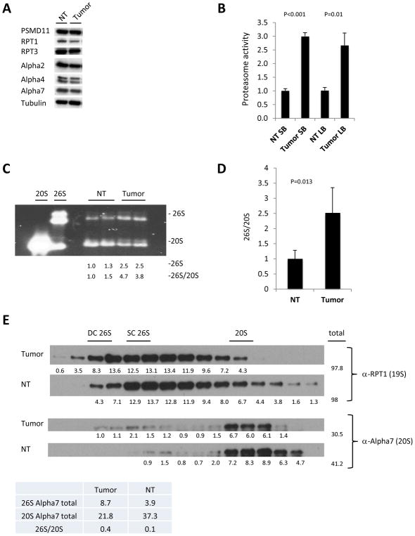
Figure 2. Enhanced 26S proteasome assembly in murine gut tumors.
A. Western blot of large bowel normal tissue and tumors from APCmin/+ mice of several individual proteasome subunits. B. Proteasome activity in normal tissue (NT) and tumors isolated from the small bowel (SB) and large bowel (LB). Values obtained for normal tissue were set as 1. The error bars represent the standard deviation from 2 biological samples per group measured in duplicates. The significance (p) between NT and tumor activity is shown. Statistical analysis was performed with a two-tailed paired t-test. C. In gel suc-LLVY-AMC activity with 0.02% SDS measured in large bowel tumors and normal tissue (NT) of AOM-DSS mice, demonstrating higher levels of 26S proteasomes and lower levels of 20S compared in tumors, indicating enhanced assembly of 26S in tumors. Densitometry was performed using ImgeJ. 26S proteasome assembly status(26S/20S) is reported as the intensity of 26S divided by intensity of 20S. D. Pooled densitometric quantitation of native gels of APCmin/+ mice normal tissue and tumors from three independent experiments, 26S proteasome assembly status (26S/20S) is reported as the intensity of 26S divided by intensity of 20S. The error bars represent the standard deviation from measurements of 4 biological samples. Statistical analysis was performed with a t-test. E. Western blot analysis of fractions from size exclusion chromotography of tumor and normal tissue (NT). Blotting with α-RPT1 antibody shows shift towards larger proteasomal complexes in the tumors while blotting with α-Alpha7 antibody reveals more doubly and singly capped proteasomes species at the expense of unassembled 20S in the tumors. DC 26S – Double capped 26S, SC 26S – Single capped 26S. Densitometry was performed using ImgeJ. Table below the blots shows total of 26S and 20S peaks detected with anti-Alpha7 antibody.

