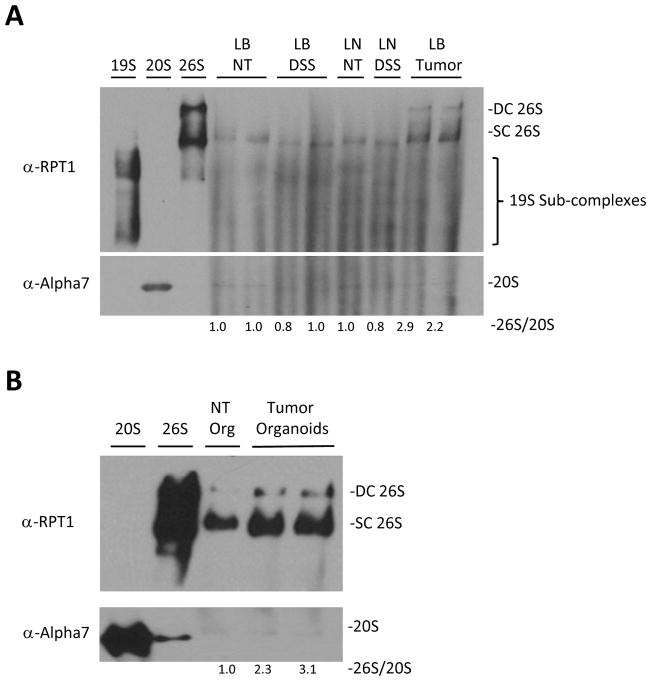
Figure 3. Enhanced assembly of 26S proteasomes, exclusively in the tumors and not other rapidly dividing cells.
A. Western blot analysis following native gel electrophoresis of large bowel tumors of APCmin/+ mice (LB Tumor), large bowel epithelium of their littermate controls (LB NT), large intestinal epithelium of DSS induced colitis mice (LB DSS) and mesenteric lymph nodes of WT (LN) and DSS treated mice (LN DSS). It demonstrates high level of 26S proteasomes exclusively in the tumors irrespectively of inflammation. B. Western blot analysis following native gel electrophoresis of organoids from large bowel normal tissue and tumors of APCmin/+ mice shows increase in the assembly of 26S protesomes in tumor organoids compared to normal tissue. 26S and 20S were detected by anti-RPT1 and anti-Alpha7 antibodies respectively. DC 26S – Double capped 26S, SC 26S – Single capped 26S. Densitometry was performed using ImgeJ. 26S proteasome assembly status(26S/20S) is reported as the intensity of 26S (DC+SC) divided by intensity of 20S.

