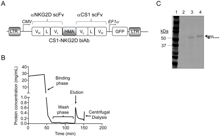
Figure 2. Design and purification of CS1-NKG2D biAb by metal-affinity chromatography.
(A) Schematic diagram of the lentiviral construct for mammalian expression of CS1-NKG2D biAb in CHO-S cells. (B) A typical profile of the protein eluted from immobilized metal-affinity chromatography column using stepwise imidazole gradient. (C) SDS-PAGE for eluted protein. Lane 1: molecular weight marker (kDa); Lane 2: protein lysate from mock transduced CHO-S cells; Lane 3: eluted control biAb (dashed arrow); Lane 4: eluted CS1-NKG2D biAb (solid arrow). Results shown are representative of at least ten independent experiments.

