Figure 3.
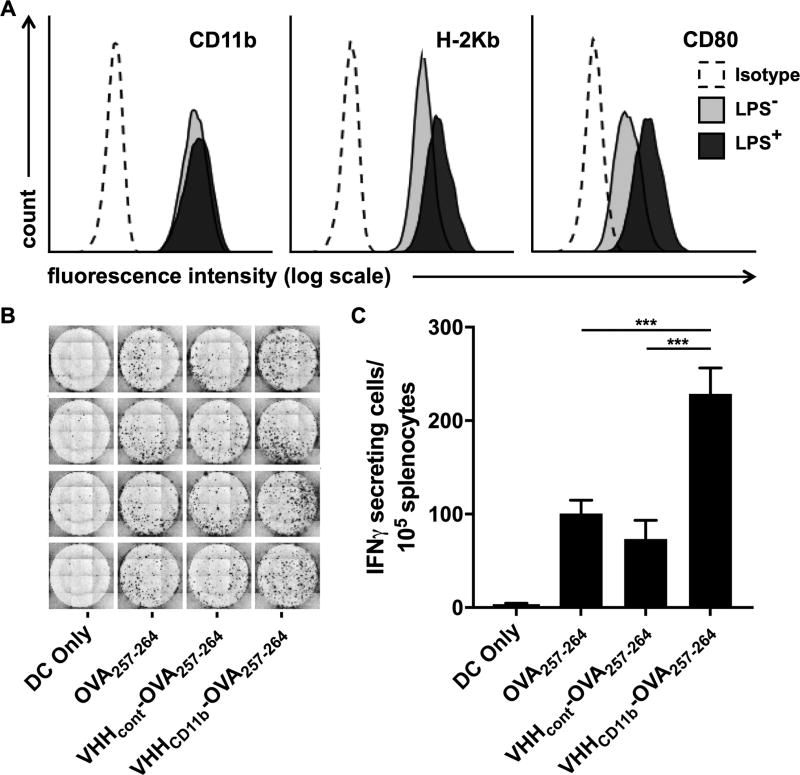
Antigen presentation is increased with VHHCD11b. A) DC2.4 cells express CD11b, H-2Kb (MHCI known to present OVA257–264), and CD80 (costimulatory molecule) as assessed via cytofluorimetry (light gray histograms). Isotype controls (Isotype) are shown as empty dashed histograms. CD80 and H-2Kb expression were increased following 24-hr incubation with LPS (dark gray histograms). B-C) DC2.4 cells were treated as indicated before LPS stimulation, followed by coculture with splenocytes from a RAG−/− OT-I mouse (1 DC2.4 cell: 20 OT-I splenocytes), and the number of IFNγ-secreting cells was measured via ELISpot assay. The spots shown in B are quantified in C (means ± SD are shown; ***P < 0.001) of an experiment performed in quadruplicate, and is a representative to three independent experiments.

