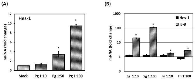
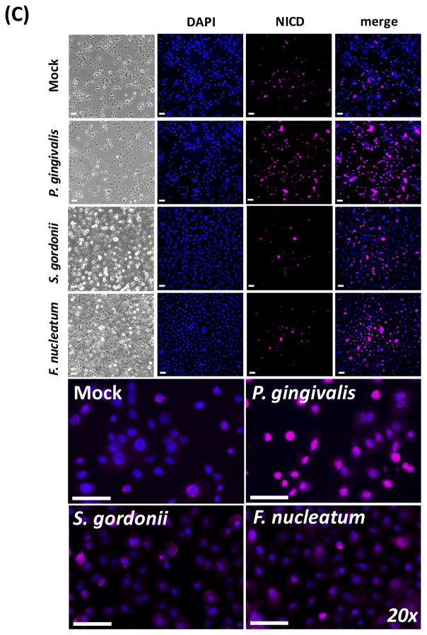
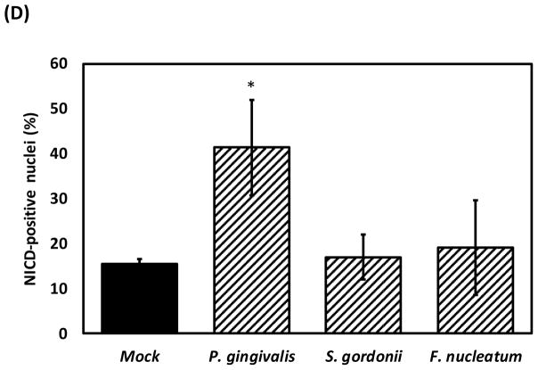
Figure 3. Notch-1 receptor is activated by P. gingivalis in oral epithelial cells.
Notch-1 activation was evaluated by quantifying mRNA levels of Hes-1 by qRT-PCR in OKF6 cells exposed to (A) P. gingivalis or (B) F. nucleatum (Fn) and S. gordonii (Sg) for 48h. IL-8 expression was used as a control. (C) Nuclear levels of Notch-1 intracellular domain (NICD) in OKF6 cells exposed or not to bacteria, were evaluated and quantified by immunofluorescence as described in methods. Micrographs are showing mock-treated (top) and bacteria-treated OKF6 cells (Bottom) for bright field, DAPI, NICD and DAPI/NICD merge. Higher magnification (20X) of DAPI-NICD merged images is also shown. Scale = 50μm. (D) Percentage of cells positive for nuclear NICD from panel (C) was determined as described in methods. The mean ± SD of triplicates from each treatment group from a representative experiment of at least 2 independent experiments are shown.*p≤0.01 when bacteria-challenged cells were compared with un-stimulated (Mock) cells.


