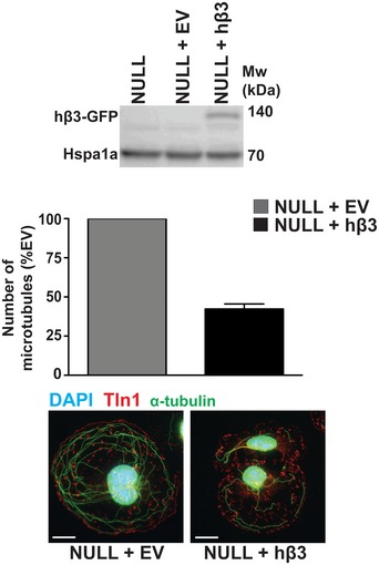
Figure EV3. Measuring the effects on microtubule stability of reintroducing β3‐integrin into β3NULL ECs.

Top: β3NULL endothelial cells were transfected with a full‐length human β3‐integrin (hβ3) cDNA expression construct or an empty vector (EV) control and Western‐blotted for β3‐integrin (β3NULL parent cells shown for comparison). Bottom: β3NULL + EV or β3NULL + hβ3 endothelial cells were adhered to fibronectin‐coated coverslips for 75 min at 37°C before being moved to ice for 15 min. Soluble tubulin was then washed out using PEM buffer (see Materials and Methods) before fixing with −20°C methanol. Immunostaining was carried out for α‐tubulin (green) and talin‐1 (Tln1‐red). DAPI (blue) was used as a nuclear stain. Images shown are representative of the data shown in the bar graph above. Bars = mean (±SEM) number of cold‐stable microtubules per cell (n = 96 cells per genotype). Scale bar = 5 μm.
