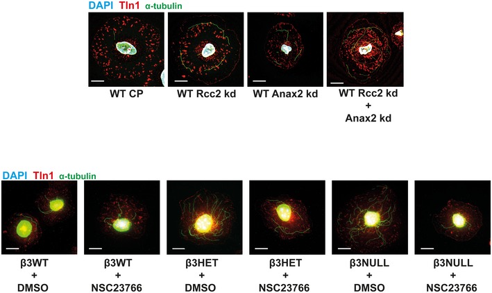
Figure EV4. Representative microtubule staining in siRNA‐treated and NSC23766‐treated ECs.

Top: β3WT ECs were transfected with control pool (CP), Anxa2 smart pool siRNA, Rcc2 smart pool siRNA or both, and allowed to recover for 48 h. They were then adhered to fibronectin‐coated coverslips for 75 min at 37°C before being moved to ice for 15 min. Soluble tubulin was then washed out using PEM buffer before fixing with −20°C methanol. Immunostaining was carried out for α‐tubulin (green) and talin‐1 (Tln1‐red). DAPI (blue) was used as a nuclear stain. Scale bar = 5 μm. Bottom: β3WT, β3HET and β3NULL cells were adhered to fibronectin‐coated coverslips for 60 min at 37°C before treated with DMSO or 50 μM NSC23766 and incubated at 37°C for a further 15 min. Cells were then moved to ice for 15 min. Soluble tubulin was washed out using PEM buffer before fixing with −20°C methanol. Immunostaining was carried out for α‐tubulin (green) and talin‐1 (Tln1‐red). DAPI (blue) was used as a nuclear stain. Scale bar = 5 μm.
