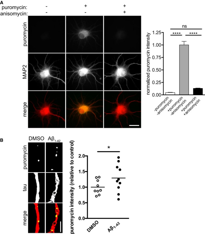
Figure EV1. Validation of puromycylation assay.

- Hippocampal neurons were cultured for 10–11 DIV. Puromycin was added to dissociated hippocampal neurons in the presence of vehicle or anisomycin. The neurons were fixed and immunostained for puromycin and MAP2. Mean ± SEM of 25–30 optical fields per condition (n = 3 independently performed experiments per group). ****P < 0.0001; ns, not significant; one‐way ANOVA with Bonferroni's multiple comparisons test. Scale bar, 10 μm.
- Hippocampal neurons were cultured for 10–11 DIV. Aβ1–42 was added to the cultured for 30 min, and puromycin was added 10 min prior to fixation. Neurons were fixed and immunostained for puromycin and tau. Mean of 9–10 optical fields per condition (sampled from two coverslips per condition) from one primary culture. *P < 0.05; unpaired, one‐tailed t‐test. Scale bar, 5 μm.
