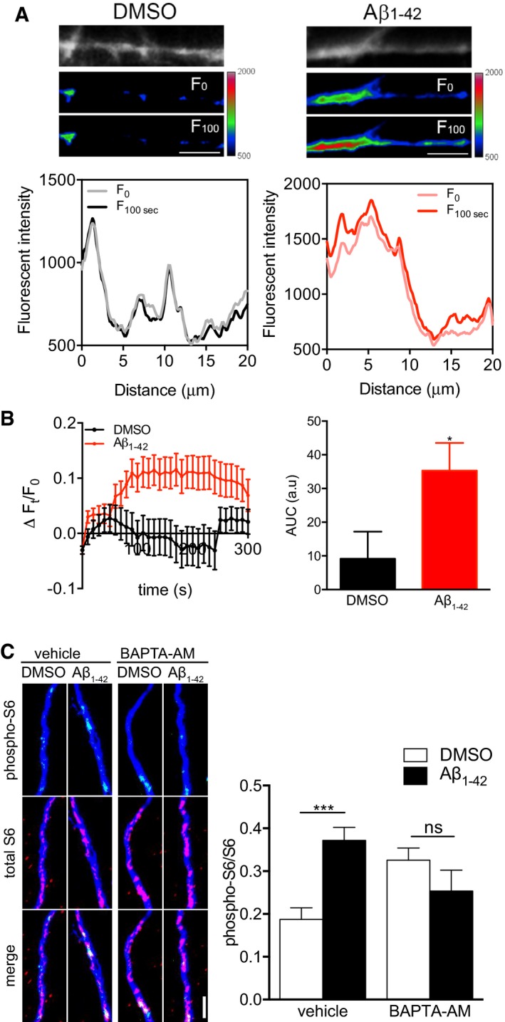
Figure 2. Immediate Aβ1–42‐dependent S6 activation requires Ca2+ signaling.

- Axons of hippocampal neurons grown in microfluidic chambers for ≥ 10 DIV were loaded with Fluo4‐AM, treated with DMSO or Aβ1–42, and live‐imaged for 5 min at 10‐s interval. Images of the Fluo4‐AM signal in representative axonal segments are shown in grayscale and pseudocolor for DMSO‐ (left) and Aβ1–42‐treated (right) axons at t = 0 s and t = 100 s. The fluorescence profile along the axonal segments at t = 0 s and t = 100 s is shown. Scale bars, 5 μm.
- Time course of the Fluo4‐AM signal (left) and cumulative fluorescence Fluo4‐AM intensity in area under curve (AUC) (right) of DMSO and Aβ1–42‐treated axons. Baseline correction was applied. Mean ± SEM of 55–65 individual axons (11–13 chambers per group; n = 6 independently performed experiments). *P < 0.05; unpaired t‐test.
- Hippocampal neurons were cultured in microfluidic chambers for 11–12 DIV, and axons were treated with vehicle or BAPTA‐AM (10 μM) for 30 min. Axons were then treated with vehicle or Aβ1–42 for 30 min and immunostained for phospho‐S6, total S6, and βIII‐tubulin. Mean ± SEM of 25 optical fields per condition (n = 5 independently performed experiments). ***P < 0.001; ns, not significant; two‐way ANOVA with Bonferroni's multiple comparisons test. Scale bar, 5 μm.
