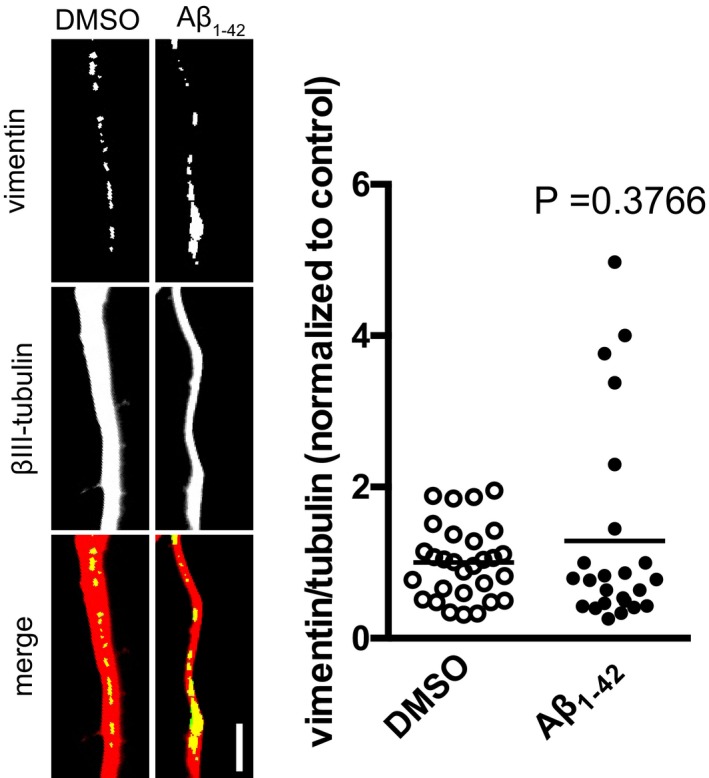
Figure EV3. Effect of 15‐min treatment on axonal vimentin levels.

Hippocampal neurons were cultured in microfluidic chambers for 11–12 DIV, and axons were treated with vehicle or Aβ1–42 for 15 min. Axons were immunostained for vimentin and βIII‐tubulin, and the vimentin signal was normalized to the βIII‐tubulin signal. Mean of 24–29 optical fields per condition (sampled from 5 to 6 coverslips per condition) from one primary culture. Unpaired t‐test. Scale bar, 5 μm.
