
Figure 6. Map7 moves toward the MT plus‐end during in migrating cells.
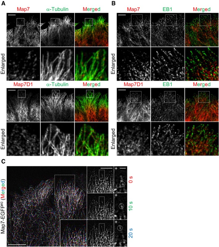
- Subcellular localization of endogenous Map7 or Map7D1 at the leading edge during cell migration. Fixed cells were co‐immunostained for α‐tubulin.
- Subcellular localization of endogenous Map7 or Map7D1 co‐stained for EB1 at the leading edge. Cells were fixed 1 h after wounding and then stained with the indicated antibodies. Enlarged merged images show that Map7/7D1 was not co‐localized with EB1.
- Live imaging of Map7‐EGFPKI cells. Time‐lapse images were taken at 10‐s intervals after wounding. Images at individual time points are shown at right, and the merged image is shown at left. Note that a Map7‐EGFPKI dot moves toward the cell cortex, where MT plus‐ends are enriched (circles).