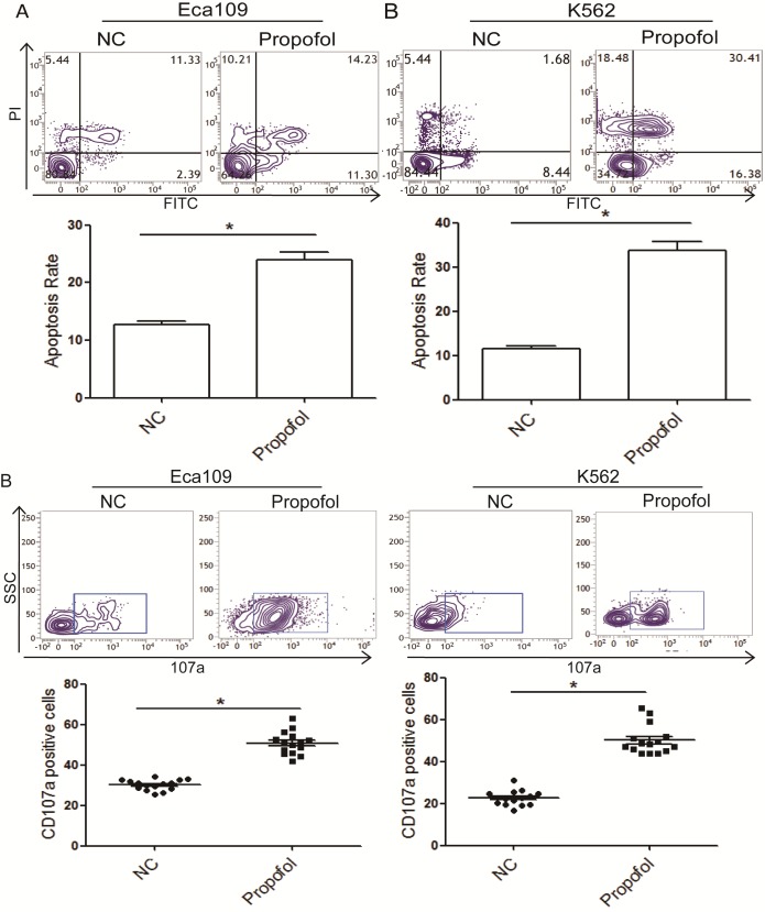
Figure 8.
Propofol enhances the cytotoxicity of NK cells to K562 and Eca109 cells, respectively. (A) Representative flow cytometry images and quantitative analysis of the apoptosis rate of K562 and Eca109 cells treated with propofol. (B) Representative flow cytometry image for CD107a positive rate analysis for K562 and Eca109 cells cocultured with propofol. *P<0.05. NK, natural killer; ESCC, esophageal squamous cell carcinoma; CD, cluster of differentiation; PI, propidium iodide; NC, negative control; FITC, fluorescein isothiocyanate; SSC, side-scattered light.

