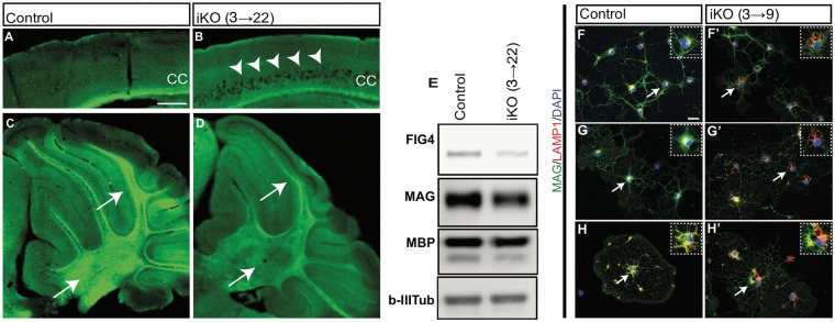
Figure 1.
Global neonatal inducible ablation of Fig4 leads to CNS degeneration, hypomyelination, and perinuclear accumulation of LAMP1+ vacuoles. (A, B) Sagittal sections of the P22 neocortex of Fig4 control (Fig4+/+ or Fig4+/flox) and Fig4 iKO(3→22) mice stained with Fluoromyelin Green. In the Fig4 iKO(3→22) neocortex, arrowheads point to extensive spongiform degeneration in deep cortical layers. Myelin staining in the corpus callosum (CC) is reduced in iKO mice. (C, D) Sagittal sections of Fig4 control and Fig4 iKO(3→22) cerebellum. Arrows in D point to cerebellar lobules and deep cerebellar nuclei with reduced Fluoromyelin staining. Scale bar, A–D = 200 μm; n = 2 mice per genotype. (E) Western blots of cerebellar lysates prepared from Fig4 control (n = 2) and Fig4 iKO(3→22) mice (n = 2), probed with antibodies specific for FIG4, myelin-associated glycoprotein (MAG) and myelin basic protein (MBP). Class III β-tubulin, detected with the TuJ1 antibody, is shown as a loading control. (F–H’) Representative images of primary OLs isolated from Fig4 control and Fig4 iKO(3→9) pups by immuno-panning with (F, F’) anti-PDGFRα to capture OPCs, (G, G’) the O4 antibody to capture immature OLs and (H, H’) anti-MOG to capture myelinating OL. After four days in vitro, cultures were fixed and stained with anti-MAG (green), anti-LAMP1 (red) and the nuclear dye DAPI (blue). LAMP1+ structures in OLs are marked by arrows and shown at higher magnification in inserts. Scale bar, F–H’ = 20 μm.

