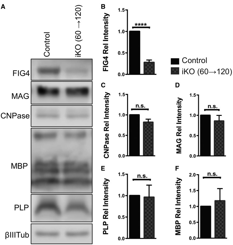
Figure 4.
Myelin abundance is not affected by adult inducible Fig4 deletion after 60 days. (A) Representative Western blots of brain membranes isolated from adult Fig4 control and Fig4 iKO(60→120) mice, probed with antibodies specific for FIG4 and the myelin proteins MAG, CNPase, MBP and PLP. Neuronal classIII β-tubulin is shown as loading control. (B–F) Quantification of FIG4 and myelin protein signals normalized to classIII β-tubulin. Relative protein intensities compared to Fig4 control brains are shown as mean value ± SEM. N = 4 per genotype. Unpaired Student t-test, ****P < 0.0001; n.s. = not significant.

