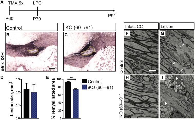
Figure 6.
In the adult CNS Fig4 is required for white matter repair. (A) Timeline showing tamoxifen administration, five consecutive injections at P60-65, stereotaxic injection of LPC in the corpus callosum (P70) and animal analysis (P91). (B, C) Coronal sections through the lesion center of adult Fig4 control and iKO(60→91) mice stained for Mbp transcript. The white dashed line demarcates the lesion area in the corpus callosum. The dashed yellow line indicates the non-myelinated area in the lesion core. Scale bar = 200μm. Quantification of the percent remyelination (white area − yellow area)/(white area), reveled less complete myelination in iKO mic; n = 6 (control) and n = 8 (iKO). (F, H) Electron photomicrographs through the corpus callosum of Fig4 control and iKO(60→91) mice. (G, I) Electron photomicrographs through the lesion core of Fig4 control and iKO(60→91) mice. Arrows in G point to re-myelinated axons. Fig4 iKO show extensive vacuolization in the lesion area. Scale bar, 2 μm. N = 4 mice per genotype.

