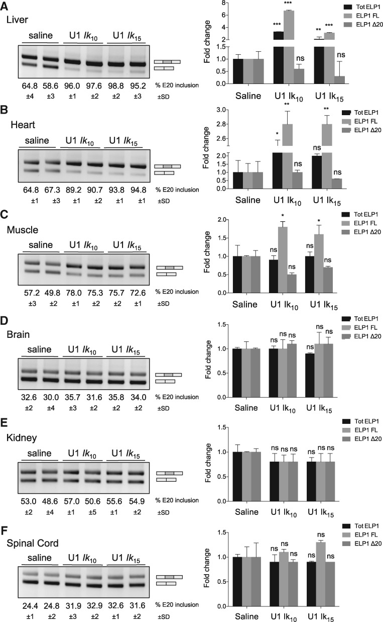
Figure 5.
AAV9-mediated delivery of ExSpeU1s Ik10 and Ik15 rescues the hELP1 splicing defect in FD mouse model. (A–F) Endpoint (gels) and quantitative (histogram) PCR analysis of hELP1 mRNA isoforms in tissues of transgenic FD mice treated with 3.55×10¹¹ and 2.65×10¹¹ viral genomes/mouse of AAV9-ExSpeU1 Ik10 (U1 Ik10) and AAV9-ExSpeU1 Ik15 (U1 Ik15), respectively. The upper band of 202 bp corresponds to exon 20 inclusion and the lower band of 128 bp corresponds to exon 20 skipping. The percentage of exon inclusion is expressed as mean±SD of groups of four animals per condition. The histograms on the right represent the fold change of ELP1 mRNAs’ isoforms (total ELP1, ELP1 full-length and ELP1 Δ20) compared to four control animals per condition. Statistical analysis was performed by two-way ANOVA (***P<0.0001, **P<0.001, *P<0.01; ns, not significant).

