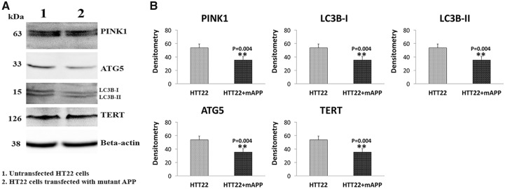
Figure 4.
Immunoblotting analysis of autophagy and mitophagy proteins in mutant APP-HT22 cells. Figure 4A shows representative immunoblotting analysis of HT22 cells transfected and untransfected with mutant APP cDNA. Figure 4B shows quantitative densitometry analysis of autophagy proteins. Autophagy proteins ATG5 (P=0.004), LC3BI (P=0.004), LC3BII (P=0.004) and TERT (P=0.004) and mitophagy protein PINK (P=0.04) were significantly decreased in HT22 cells transfected with mutant APP cDNA relative to untransfected HT22 cells.

