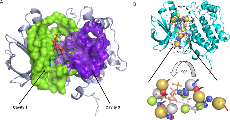
Figure 2.
Detected cavities and a pharmacophore model for Polo-like kinase 1 (PDB ID: 2OU7) shown in Pymol. (A) Two cavities detected by CAVITY. Green: active site; purple: potential allosteric site. (B) The pharmacophore model of the active ATP binding site. Blue and red arrows represents the H-bond donor center and h-bond acceptor, respectively; green spheres: hydrophobic center; olive spheres: positive electrostatic center; grey spheres: negative electrostatic center.

