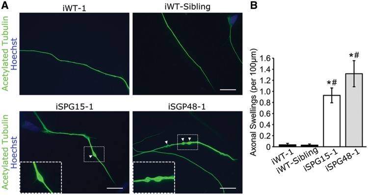
Figure 2.
Increased neurite swellings within SPG15 and SPG48 telencephalic neurons. (A) Representative images of telencephalic neurons with axonal swellings labeled with arrowheads. Boxed regions are enlarged in the insets. Scale bars: 20 µm. (B) Quantification revealed a significant increase in axonal swellings in patient-derived neurons compared with control neurons. Data are presented as means ± SEM from at least 30 cells in three independent experiments. *P < 0.05 compared with iWT-1. #P < 0.05 compared with iWT-sibling cells.

