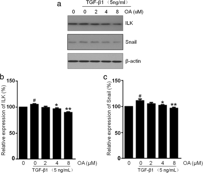
Fig. 6.
Effects of OA on the ILK and Snail expression in NRK-52E cells. The cells were incubated with 5 ng/mL of TGF-β1 for 48 h with different concentrations of OA (0, 2, 4, 8 μM). a The expression level of ILK and Snail was determined by Western blotting. b, c The expression level was quantitatively analyzed with Image J software. The data showing mean ± SD. # P < 0.05 vs. 0 ng/mL TGF-β1. * P < 0.05, and ** P < 0.01 vs. 0 μM OA in the presence of 5 ng/mL TGF-β1

