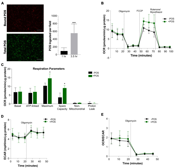
Figure 4.
Increased maximum respiration and spare capacity of hfRPE cells exposed to bovine POS. A: Representative 20X field images of bound (red) and total (green) POS at 3.5 h, and quantification of ingested photoreceptor outer segments (POS) from six images per time point at 1.0 and 3.5 h compared with an unpaired t test. B: Mitochondrial stress tests were performed on paired samples from four different human fetal RPE (hfRPE) cell lines challenged (+POS) or unchallenged (-POS) with bovine POS 3.5 h before testing (green and black, respectively). C: Respiration parameters were calculated from the mitochondrial stress test measurements. D and E: Basal extracellular acidification rate (ECAR) values obtained during the mitochondrial stress test revealed no obvious differences between the POS-treated and untreated samples or in the oxygen consumption rate (OCR)/ECAR ratios. Technical triplicates were conducted for each condition for each line. *p<0.05, *** p<0.001. Results are expressed as mean ± standard deviation.

