Fig. 5.
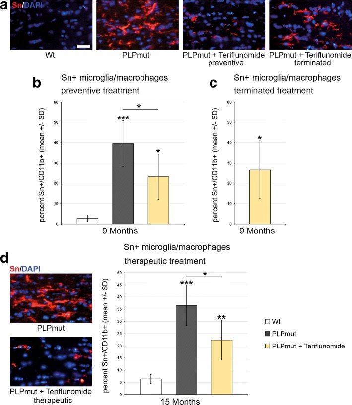
Teriflunomide treatment significantly impairs microglial activation in optic nerves of PLPmut mice. a Representative immune fluorescence microscopy of Sn+ microglial cells in longitudinal optic nerve sections from Wt, untreated mutants (PLPmut), preventively treated mutants (150 days, starting from postnatal month 4; PLPmut + teriflunomide preventive) and PLPmut mice after treatment interruption at 75 days after treatment onset (PLPmut + teriflunomide terminated). Mice were investigated at 9 months of age. b Quantification of Sn + microglial cells in optic nerve sections of Wt and PLPmut mice and in PLPmut mice after 150 days of preventive treatment. The numbers of Sn + activated microglial cells were significantly increased in the untreated PLPmut mice, which was partially forstalled upon preventive treatment. Mice were investigated at 9 months of age. c Quantification of Sn + microglial cells in optic nerve sections of PLPmut mice after 75 days of preventive treatment, followed by 75 days without treatment. Reduced elevation of Sn+ microglial cells after terminated treatment was not significant anymore. Mice were investigated at 9 months of age. d Immunofluorescent depiction of Sn+ microglial cells and their quantification (right) in optic nerve sections of Wt and PLPmut mice and in PLPmut mice after 150 days of therapeutic treatment (PLPmut + teriflunomide therapeutic) starting at 10 months of age. Therapeutic treatment significantly reduced the elevation of Sn+ microglial cell numbers in PLPmut mice. Mice were investigated at 15 months of age. Scale bar, 30 μm. One-way ANOVA and Tukey’s post hoc tests. *P < 0.05, **P < 0.01, ***P < 0.001. n = 5 mice per group

