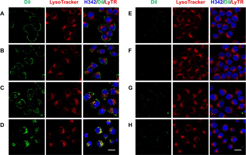
Figure 2.

Live-cell tracking of DiI-labeled LP-9AzSia. FR+ HeLa cells were incubated with f/DiI-LP-9AzSia (A-D) or DiI-LP-9AzSia (E-H) at the 9AzSia-based concentration of 100 μM for 0.5 h. The cells were changed into fresh medium and imaged by confocal fluorescence microscoy at varied time points including 1.5 h (A, E), 3 h (B, F), 6 h (C, G) and 24 h (D, H). The liposomes were tracked with DiI (green), the lysosomes were stained with LysoTracker Deep Red (red), and the nuclei were stained with Hoechst 33342 (blue). The intensity of DiI at 24 h (D, H) was adjusted to 50% for the illustration purpose. Scale bar, 20 μm.
