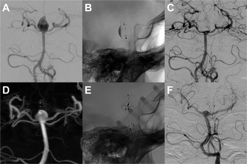
Figure 2.
Ruptured aneurysm (A) of the basilar apex that was treated with a WEB DL device in the acute phase. The profile projection in (B) depicts the device with contrast stasis in the mesh. The final result was a small neck remnant (C). A constantly growing aneurysm recurrence. (D) was treated 40 months later using a WEB SL device (E). For protection of the right P1 segment a self-expanding stent (Enterprise 4×16 mm) was placed. Follow-up after 7 months shows a small neck remnant (F).

