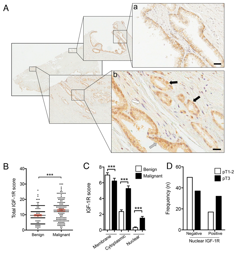
Figure 1. Nuclear IGF1R is associated with advanced tumor stage.
A. IGF-1R IHC in radical prostatectomy: a) Benign epithelium showing membrane IGF-1R, with cytoplasmic IGF-1R in basal cells; b) Mixed Gleason 3 (grey arrow) and 4 (black arrow) cancer containing more IGF-1R than benign epithelium, prominent cytoplasmic and nuclear IGF-1R, and perineural invasion (white arrow). Scale bar 20μm. B.IGF-1R IHC scored for total IGF-1R (n= 137 RPs). Graph: total IGF-1R score (bars, mean ± SEM, in red) in benign and malignant epithelia. The cancers contained significantly more IGF-1R than benign prostatic epithelium from the same RP (***p=0.001, Wilcoxon matched pairs signed rank test).C. IGF-1R quantification in plasma membrane, cytoplasm, nucleus (n=137 RPs, ***p<0.001, Wilcoxon test). D. Stage pT3 prostate cancers contain more nuclear IGF-1R than stage pT1-2 cancers (p=0.011).

