
Fig. 2.
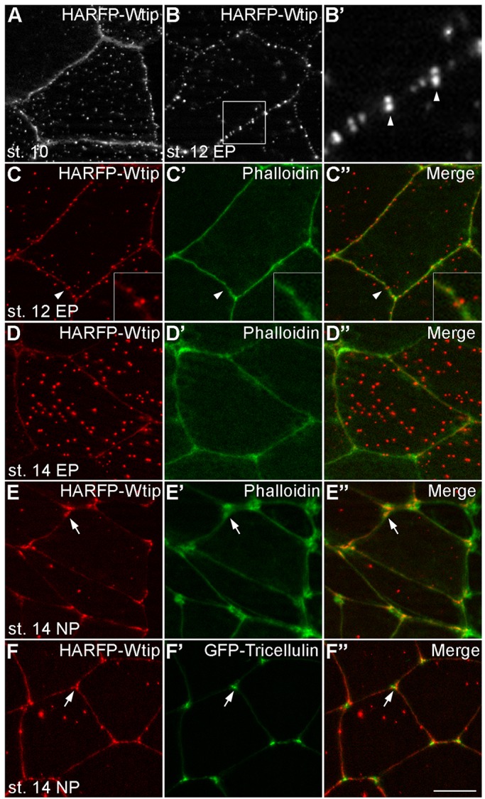
Dynamic subcellular localization of Wtip in Xenopus embryonic ectoderm. HA-RFP-Wtip RNA (200 pg) was injected into animal blastomeres of Xenopus four-cell embryos. The injected embryos were fixed at different stages and superficial ectoderm was imaged. (A) Stage 10 gastrula ectoderm; (B,B′) stage 12 epidermal ectoderm. The boxed area in B is enlarged in B′. Note the punctate distribution of Wtip along the cell junctions, with some puncta paired across the junction (arrowheads). (C-E″) Stage 12 (C-C″) or stage 14 (D-D″) epidermal ectoderm or stage 14 neuroectoderm (E-E″) stained with Phalloidin to visualize F-actin at the cell junctions. Arrowheads indicate a pair of Wtip puncta on the opposite sides of the junction, which is enlarged in the inset. Note the presence of Wtip at tricellular junctions (arrows). (F-F″) Stage 14 neuroectoderm coinjected with HA-RFP-Wtip and GFP-Tricellulin (50 pg) RNAs. The Wtip signal surrounds Tricellulin at tricellular junctions (arrows). Scale bar: 10 µm. Representative images from three to five independent experiments are shown, and each group included explants from 8-12 embryos.