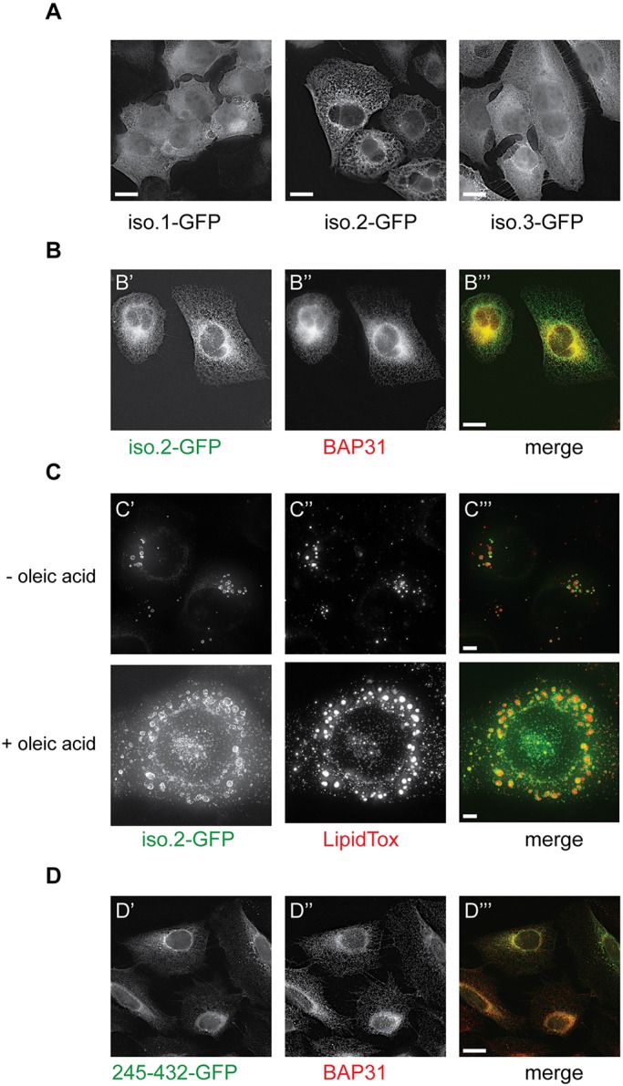
Fig. 2.

USP35 isoforms localise to distinct intracellular compartments. (A) U2OS FlpIn inducible cells expressing USP35 isoforms bearing a C-terminal GFP tag were fixed with paraformaldehyde (PFA) and stained with anti-GFP antibody. Protein localisation was examined by fluorescence microscopy. (B) As in A, but samples were simultaneously stained with anti-GFP and anti-BAP31 (an ER marker) antibodies. (C) U2OS FlpIn USP35iso2 cell line was left untreated or loaded with 0.3 mM oleic acid for 16 h at which point expression of isoform 2 was induced. Following an 8 h incubation, cells were fixed with PFA and stained with LipidTox Deep Red. LipidTox staining and intrinsic GFP fluorescence was analysed by microscopy. (D) Expression of USP35245-432 with a C-terminal GFP tag in stably transfected U2OS FlpIn cells was induced with tetracycline. Cells were fixed, stained with anti-GFP and anti-BAP31 antibodies, and samples analysed by immunofluorescence microscopy. Scale bars: 15 µm (A,B,D), 5 µm (C).
