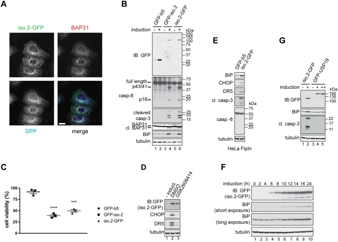
Fig. 4.
USP35 isoform 2 overexpression induces ER stress and apoptosis. (A) Expression of USP35iso2 tagged at the C-terminus with GFP was induced in U2OS FlpIn cells with tetracycline for 48 h. Cells were fixed with PFA, co-stained with anti-GFP, anti-BAP31 and anti-SPP (signal peptide peptidase) antibodies, and analysed by microscopy. Scale bar: 15 µm. (B) Expression of USP35iso2 or a control ER-localised construct GFP-b5 was initiated by adding tetracycline to U2OS FlpIn cell lines. After 48 h, cells were lysed, and samples resolved by SDS-PAGE and immunoblotted (IB) using antibodies against the indicated proteins. cl. BAP31, cleaved BAP31. p43, p41 and p18, cleaved products of pro-caspase 8. (C) As in B, but the cell lines were seeded into a 96-well plate and, at 48 h post-induction, an MTS proliferation assay was performed. The values on the y-axis represent the ratio of absorbance read for induced to non-induced cells. Error bars indicate s.e.m. (n=3 biological replicates). ****P<0.0001, ***P=0.0002 (one-way ANOVA). (D) USP35iso2 tagged at the C-terminus with GFP was expressed in the inducible U2OS FlpIn cell line for 24 h in the presence of 5 µM GSK2606414 or with DMSO (control). Cells were lysed, and samples resolved by SDS-PAGE and immunoblotted using antibodies against the indicated proteins. (E) As in B, but HeLa FlpIn cell lines induced to express GFP-b5 or USP35iso2 were used. (F) Expression of USP35iso2 bearing a C-terminal GFP tag was induced in U2OS FlpIn cell line. Levels of the GFP-tagged protein and upregulation of BiP were monitored at the indicated time points by immunoblotting cell lysates with antibodies against the indicated proteins. (G) As in B, but U2OS FlpIn cells stably transfected with GFP–USP19 were used as a control. ++ indicates a higher concentration of tetracycline used for GFP–USP19 expression.

