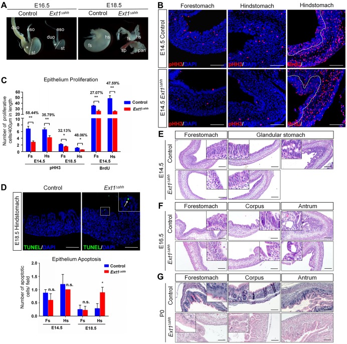
Fig. 2.
Depletion of epithelial HS causes stomach growth defects and impaired gastric morphogenesis. (A) Gross images of stomachs at indicated embryonic stages from control and Ext1Δshh mutant mice. Mutant embryos exhibit smaller stomachs and thinner intestines. Asterisks represent the glandular stomachs. Dashed lines separate the forestomach and hindstomach. Note that abnormal and rudimentary ‘buds’ are observed from some E18.5 mutant embryos. eso, esophagus; duo, duodenum; st, stomach; fs, forestomach; hs, hindstomach; sp, spleen; pan, pancreas. (B,C) Analysis of gastric epithelium proliferation by pHH3 and BrdU immunostaining. The mutant stomach shows a decreased proliferation in both the forestomach and hindstomach at E14.5. Quantification is achieved by counting the number of pHH3- and BrdU-positive cells relative to the length of the epithelium. The rates of reduction between control and mutant are shown. Nuclei are visualized with DAPI. Dashed lines outline the epithelium. n=5 per group. (D) Increased apoptosis at E18.5 is observed in mutant glandular stomach, demonstrated by TUNEL assay. The inset represents magnified image of the boxed area. The arrow indicates an apoptotic cell in the mutant epithelium. n=3 per group. (E-G) Hematoxylin and Eosin stained sections of mutant stomachs showing histological defects during gastric epithelium morphogenesis. *P<0.05, **P<0.01; n.s., no significant difference. Error bars indicate s.e.m. Scale bars: 100 μm.

