
Fig. 6.
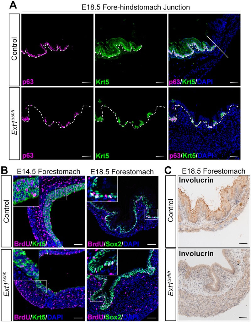
Epithelial HS regulates forestomach stratification and differentiation. (A) Immunostaining for basal cell markers on forestomach sections. The number of p63+ and Krt5+ basal cells declined and their distribution is sporadic rather than continuous in the mutant epithelium. The straight line separates the forestomach and glandular stomach. White dashed lines outline the epithelium. (B) Analysis of the proliferation in forestomach epithelium. Co-staining for BrdU and Krt5 indicates that remaining basal cells in mutant have proliferation capacity. However, those abnormal Sox2-stained cells in the supralayer are negative for BrdU staining. Magnified views of the boxed area are presented in the insets. (C) Immunohistochemistry analysis of keratinization marker involucrin reveals that the formation of squamous layers is disrupted in the mutant. Scale bars: 50 μm.