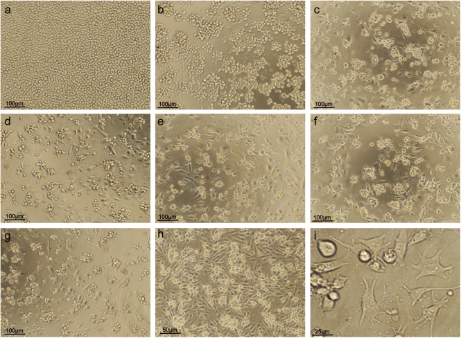
Fig. 3.
Effects of PMA concentration on differentiation of THP-1 cells to macrophage-like cells induced by PMA. (A) When no PMA was added to the THP-1 cells, the THP-1 cells floated in the cell culture solution with significant proliferation. (B) When the PMA concentration increased to 2 ng/ml, a few of the THP-1 cells adhered and differentiated into macrophage-like cells. (C-I) When the PMA concentration was equal to or more than 5 ng/ml, most of THP-1 cells adhered and differentiated into macrophage-like cells. (A) 0 (×100). (B) 2 ng/ml (×100). (C) 5 ng/ml (×100). (D) 10 ng/ml (×100). (E) 20 ng/ml (×100). (F) 50 ng/ml (×100). (G) 100 ng/ml (×100). (H) 10 ng/ml (×200). (I) 10 ng/ml (×400).

