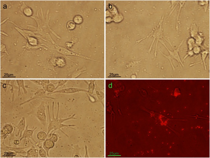
Fig. 5.
Differentiated THP-1 cells with many long pseudopodia and macrophage-like cell shape after implementing induction conditions: a lower concentration of PMA(10 ng/ml), a shorter time (72 h) and a lower density of THP-1 (0.25×10⁶/ml). (A-C) Morphology of pTHP-1 cells. (D) Phagocytosis of fluorescent beads by pTHP-1 cells.

