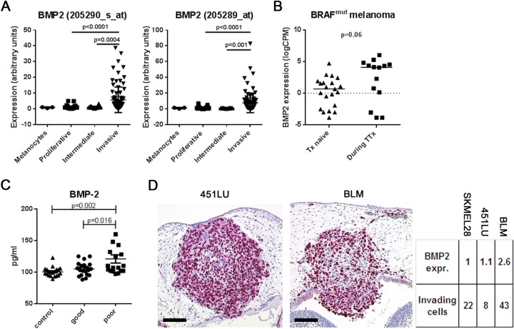
Fig. 1.
BMP-2 is up-regulated in melanoma cells with an invasive phenotype. (A) A melanoma database (http://www.jurmo.ch/php/genehunter.html) was screened for the expression level of BMP-2. In the four different datasets comprising melanocytes (n=3), proliferative (n=101), intermediate (n=26), and invasive (n=90) melanoma cell lines, BMP-2 was significantly up-regulated in melanoma cells with an invasive phenotype compared to cells with a proliferative phenotype (one-way ANOVA). (B) RNA-expression of BMP-2 in BRAF mutated patient-derived melanoma cells therapy (Tx)-naïve or during targeted therapy (TTx). A subset of melanoma cells from patients under TTx had a strong expression of BMP-2 compared to the Tx-naïve cohort (median 0.48 vs 4.10 counts per million, P=0.06, t-test). (C) BMP-2 serum levels in healthy controls (n=20) and 38 melanoma patients [n=20 stages IB-IIC (long survival), and n=18 stage IV (short survival)]. In control sera, median BMP-2 concentration was 100.5 pg/ml (89-123 pg/ml). In sera from patients with long survival it was 105.1 pg/ml (88-125 pg/ml). In sera from patients with short survival it was 120.9 pg/ml (97-201 pg/ml). The differences between controls and short-term survivors and between long-term and short-term survivors were significant (one-way ANOVA). (D) Chick embryo brain metastasis model. Histological slides were analyzed for single melanoma cell invasion in the rhombencephalon of the chick embryo and correlated with BMP-2 RNA-expression of the corresponding melanoma cell line. Expression of BMP-2 was 2.6-fold higher in highly invasive BLM compared to less invasive 451LU and SKMEL28 (SKMEL28: 22±11 invading cells per histological section, n=5 embryos analyzed; 451LU: 8±2, n=6; BLM: 43±10, n=8; P<0.05, one-way ANOVA). Scale bars: 100 µm.

