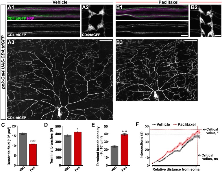
Fig. 2.
Paclitaxel treatment increases peripheral sensory dendrite branch density. (A1-B3) Confocal projections of fixed, filet-dissected vehicle- or paclitaxel-treated larvae showing class IV dendritic arborization (C4da) neuronal compartments labeled by CD4:tdGFP. (1) Individual C4da sensory axons are visible within the HRP-labeled peripheral nerve bundles. (2) C4da axon terminals project to form a ladder-like pattern in the VNC. (3) Dorsal dendrite projections of C4da ddaC shown here with cell body centered near the bottom of the frame. Axon and VNC scale bar: 10 µm; dendrite scale bar: 50 µm. (C-E) Dorsal projections of C4da dendrites labeled by CD4:tdGFP after treatment with either vehicle or paclitaxel were quantified: (C) area of dendritic field measured in ImageJ; (D) number of terminal branches counted manually; and (E) terminal branch density. Mean±s.e.m.; n=10 neurons from >5 larvae for each treatment, Student's t-test. (F) Sholl profile was performed on dorsal projections of C4da dendrites in ImageJ and plotted as the number of branch intersections with relative distance from the cell body to the anterior/posterior edges of the dendritic field. Solid lines connect mean intersections at each relative shell and shaded regions represent s.e.m.; n=10 neurons from >5 larvae for each treatment; interaction: ns, relative distance: P<0.0001, treatment group: P<0.0001, two-way ANOVA; critical value: P<0.05, critical radius: ns, Student's t-test. *P<0.05, ****P<0.0001.

