Fig. 6.
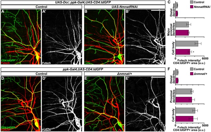
Dendritic Futsch organization does not require Nmnat. (A-B′) Confocal projections showing immunostaining of microtubule-associated protein Futsch within sensory dendrites from larvae with C4da neuronal RNAi-mediated knockdown of Nmnat (B; ppk-Gal4>UAS-Dcr,UAS-NmnatRNAi) and controls (A; ppk-Gal4>UAS-Dcr). C4da neurons exhibit continuous Futsch staining throughout the major branches, even in those with substantial dendrite loss (B). Scale bar: 50 µm. (C) Futsch intensity measured within C4da neuronal compartments, i.e. cell body, major proximal dendrite branches and distal dendrite branches, in larvae with C4da RNAi-mediated knockdown of Nmnat and controls. Mean±s.e.m.; n=5 neurons from >4 larvae from each genotype, Student's t-test. (D-E′) Confocal projections of Futsch immunostaining and C4da expression of CD4:tdGFP in Nmnat heterozygous larvae (E; Δnmnat/+) and controls. (F) Futsch intensity measured within C4da neuronal compartments in larvae heterozygous for Nmnat and controls. Mean±s.e.m.; n=5 neurons from >4 larvae from each genotype, Student's t-test. *P<0.05.

