Fig. 8.
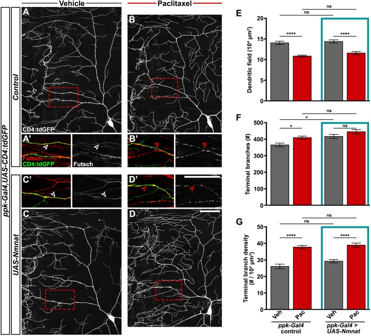
Nmnat does not prevent paclitaxel-induced dendrite density or Futsch disruption. (A-D) Confocal projections showing ddaC sensory dendrites labeled with CD4:tdGFP from control genotype treated with vehicle (A) or paclitaxel (B), and larvae overexpressing Nmnat in C4da sensory neurons (UAS-Nmnat) treated with vehicle (C) or paclitaxel (D). Scale bar: 50 µm. (A′-D′) High magnification of dendritic regions boxed in A-D, showing immunostaining of microtubule-associated Futsch in C4da dendrites (arrowheads) and other classes of peripheral neurons. Scale bar: 50 µm. (E-G) Quantification of dendritic field (E), number of terminal branches (F) and terminal branch density (G) of the dorsal projections of ddaC neurons from ppk-Gal4 control larvae and larvae overexpressing Nmnat in C4da sensory neurons after 48 h treatment with vehicle or paclitaxel. Means±s.e.m.; n=14 neurons from >7 larvae form each genotype and treatment; one-way ANOVA with Tukey's multiple comparisons test. *P<0.05, ****P<0.0001; ns, nonsignificant.

