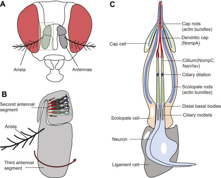
Fig. 1.
Structure of the Drosophila auditory organ. (A) A frontal view of an adult Drosophila head, with antennae marked in gray (eyes are in red). (B) Magnified image of an antenna (area highlighted in light green in A). The auditory organ (Johnston's organ) is located in the second segment of the antenna (A2) and consists of functional units called scolopidia. These are attached to the cuticle of the second antennal segment and to the A2/A3 joint. Sound particles cause the displacement of the antennal arista, which induces the third antennal segment to rotate (arrow). This acoustic, stimulus-induced rotation applies force to the cilia of mechanosensory neurons in the scolopidia, resulting in the firing of action potentials along the antennal nerve. A single scolopidium is highlighted in the light green box. Each scolopidium is attached to the antenna cuticle (arrows) by a cap cell (red) and a ligament cell (gray), as detailed in C. (C) Cellular components of an individual scolopium. Two to three mechanosensory neurons (two neurons are shown) have their ciliary dendrites enclosed by an actin-rich scolopale cell. Mechanosensory neurons express several ion channels that regulate mechanosensation, including Nan/Iav (green) and NompC (yellow). Scolopale cells form septate junctions with the ligament and cap cells at the basal and apical ends of the scolopidium, respectively, ensuring the formation of an enclosed scolopale space between the scolopale cell and neuronal cilia. The ciliary dendrites of each neuron insert into an acellular cap that contains the NompA glycoprotein (red), which is anchored to the A2/A3 joint by a cap cell. The scolopidium is attached to the A2 cuticle at its proximal end by a ligament cell. Approximately 200 scolopidia are located in Johnston's organ.

