Fig. 2.
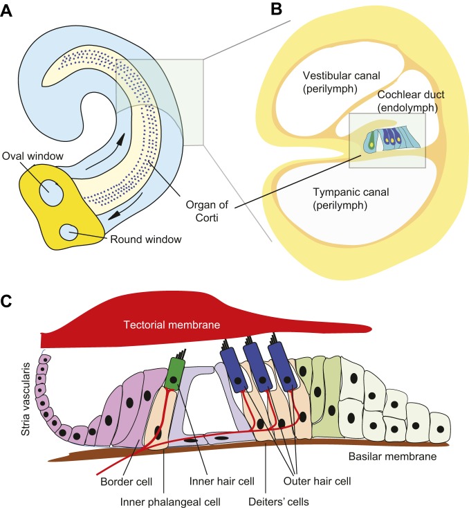
Structure of the mammalian auditory organ. (A) In the mouse inner ear, the organ of Corti is located on the basilar membrane, which runs the length of the cochlear duct. Mechanical waves conducted by the middle ear bones are transmitted to the oval window and travel through the perilymph surrounding the cochlear duct (arrows). Compression of the oval window is matched by a corresponding outward movement at the round window. (B) A cross-section of the cochlear duct, showing three rows of outer hair cells (dark blue) and one row of inner hair cells (dark green). (C) A magnified view of the arrangement of hair cells in the organ of Corti. Inner hair cells are surrounded by inner phalangeal and border cells and are separated from the outer hair cells by two pillar cells that together form the tunnel of Corti. Outer hair cells are surrounded by Deiters' cells. Inner and outer hair cells receive afferent and efferent innervation, respectively. Stereocilia located on the apical surface of hair cells are embedded into the tectorial membrane (outer hair cells) or lie just beneath the membrane (inner hair cells). During sound reception, the sound-induced vibration of the basilar membrane applies mechanical force to the tip links of the stereocilia, causing an influx of potassium and calcium and the depolarization of hair cells. The potassium gradient in the cochlear duct endolymph is maintained by the stria vascularis in the lateral wall of the cochlear duct.

