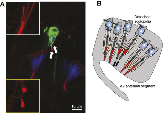
Fig. 4.
Apical detachment of scolopidia in DrosophilaUbr3 mutants. (A) A confocal image of the second antennal segment of a mosaic adult Drosophila in which GFP+ marked cells are Ubr3−/− (mutant) and GFP– cells are Ubr3+/− (wild-type control). The apical junction protein NompA is shown in red and actin (scolopale cells) in blue. Arrows indicate two detached Ubr3−/− scolopidia, which also exhibit abnormal puncta pattern of NompA. The white outline box (upper) and yellow outline box (lower) show high-power images of the NompA pattern in the apical junction from wild-type or Ubr3 mutant scolopidia, respectively. (B) A schematic summary of the apical detachment of scolopidia in Ubr3 and other Usher mutants, such as myosin VIIA and Cad99c. Arrows indicate detached scolopidia. Image in A taken from Li et al. (2016).

