Fig. 2.

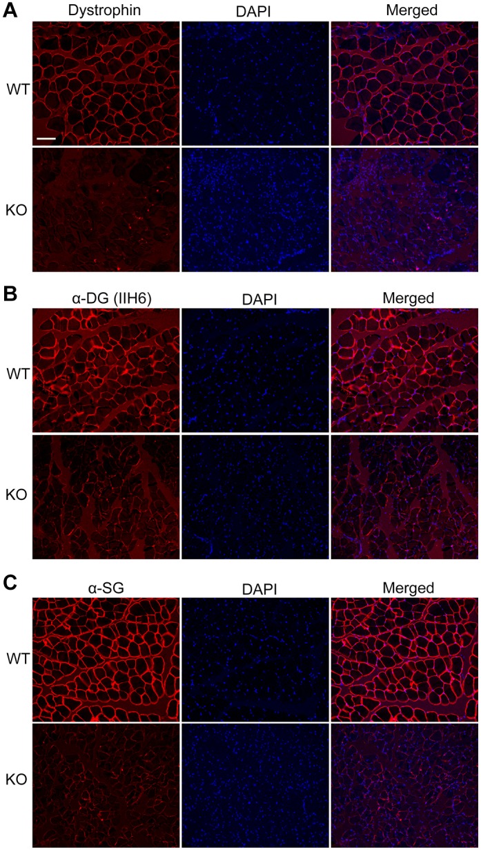
Disruption of dystrophin expression in the skeletal muscle of DMD KO rabbits. (A-C) Immunofluorescence staining of muscle sections from WT and DMD KO rabbits with mouse monoclonal antibodies against dystrophin (A), glycosylated α-dystroglycan (B) and α-sarcoglycan (C). Nuclei were stained by DAPI. Scale bar: 100 µm.
