Fig. 4.
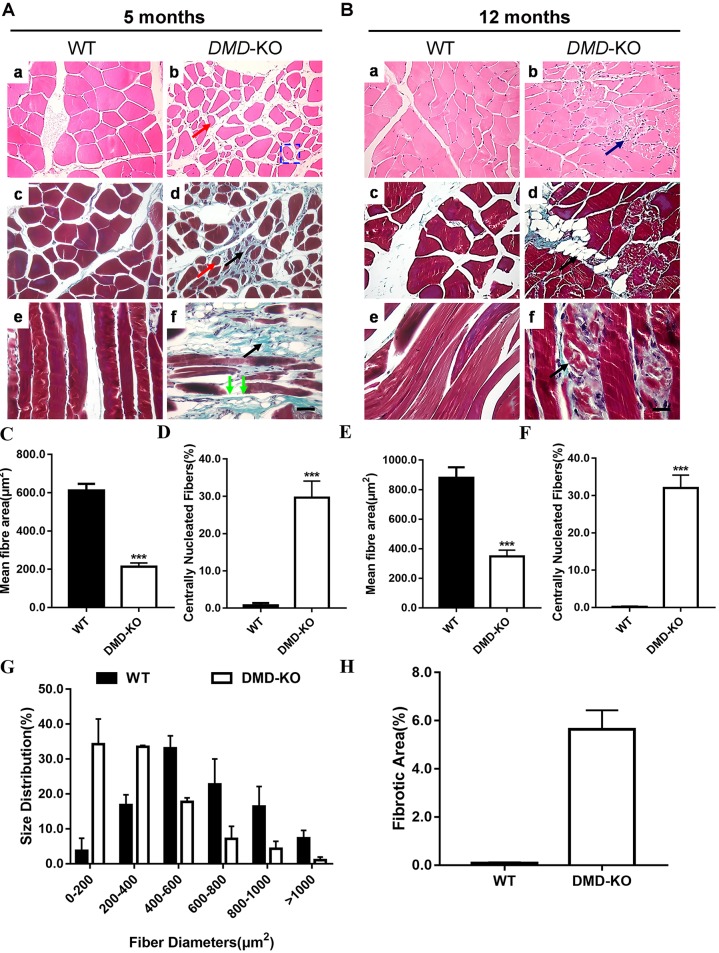
Muscular dystrophy presentation in DMD-KO rabbits. (A,B) Analysis of H&E- and Masson's trichrome-stained sections of gastrocnemius from 5-month-old (A) and 12-month-old (B) WT and DMD KO rabbits. DMD KO rabbits displayed myopathy with excessive fiber size variation (red arrows), fiber fracture (green arrows), fibrosis (black arrows) and central nucleated fibers (blue rectangle). (C,E) Quantification of mean gastrocnemius muscle fiber area in WT and DMD KO rabbits at 5 (C) and 12 (E) months of age. (D,F) Quantification of centrally nucleated fiber (CNF) percentage in WT and DMD KO rabbits at 5 (D) and 12 (F) months of age. (G) Size distribution of WT and DMD KO gastrocnemius muscle at 5 months of age. (H) Quantification of relative fibrotic area in WT and DMD KO rabbits at 5 months of age. Scale bars: 50 µm. ***P<0.001; n=5.

