
Fig. 2. Calcaneal ontogeny in apes, humans, and A. afarensis.
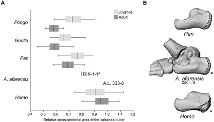
(A) Standardized cross-sectional area of the calcaneal tuber in juvenile (light gray) and adult (dark gray) apes, A. afarensis, and modern humans. The vertical line represents the taxon median, the box represents the interquartile range, and the whiskers indicate the range of the data. A higher number corresponds with increased robusticity. Notice that, unlike in apes or humans, in A. afarensis, the calcaneus exhibits a significant developmental increase in robusticity; the Dikika juvenile is chimpanzee-like, while the Hadar adult is human-like. All juveniles are significantly different from adults for each taxon (P ≤ 0.05) except for Homo sapiens (P = 0.53). (B) Three-dimensional (3D) surface renderings of DIK-1-1f, chimpanzee (top), and human (bottom) juvenile calcanei shown in lateral view. Notice the ape-like gracility of DIK-1-1f, the proximodistally long and dorsoplantarly shallow distal calcaneal region, and the large peroneal trochlea. As in humans, however, there is a plantarly positioned apophyseal flange (indicated by the asterisk) for the lateral plantar process (epiphyseal surface outlined).